
Looking into the future with experimental pathology
The CSU Experimental Pathology Facility is shaping the future of pathology by helping researchers evaluate samples and design studies that advance medical knowledge, offering clinical, anatomic, and molecular pathological expertise.
Link copied!
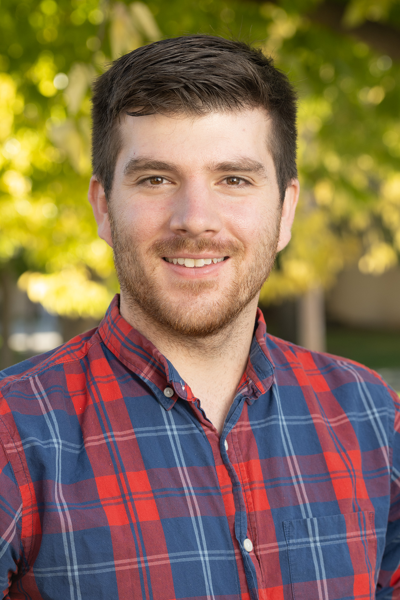
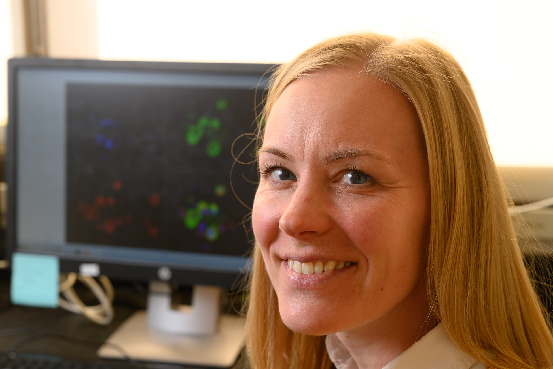
When Dr. Mac Harris, Dr. Kelly Santangelo, and lab technician Laura Ashton look at a sample slide, they are doing much more than peering into a microscope. They are helping to define the future of pathology in a rapidly changing research landscape.
Their work in the CSU Experimental Pathology Facility not only helps researchers evaluate tissue and blood samples using established techniques, it assists in designing studies that move medical knowledge forward.
“Our goal is to is to utilize pathology to bring research to the cutting edge, to be the most informative it can be,” said Harris, assistant professor in the CSU Department of the Microbiology, Immunology, and Pathology, and co-director (with Santangelo) of the lab.
The Experimental Pathology Facility was launched in 2016 by Santangelo and Dr. Brendan Podell through two consecutive “Emerging Core Facility” awards from the Office of the CSU Vice President for Research, offering clinical, anatomic, and molecular pathological expertise for both sample preparation and pathologic interpretation.
What does this mean for researchers?
“The immediate benefit is that a researcher can know that there is a service dedicated to their research study. They know their work will be prioritized and their slides will get generated in a timely manner with our histology services,” Harris said. “Another key aspect of our core is access to veterinary pathologists to help with interpretation, because it is frequently necessary to have a trained pathologist to detect the nuanced difference among lesions. And a trained pathologist can teach people about this subtle pathology and potentially unexpected findings related to their disease focus. That’s the power of working with our team.”
Housed in the Veterinary Diagnostic Laboratory on CSU’s South Campus, the core’s main mission is to provide pathological expertise to researchers within the CSU Veterinary Health System, across the CSU scientific community, and throughout the country.
“The true purpose of the Experimental Pathology Facility is to ensure that we’re documenting any pathology that’s occurring within an animal model, whether it’s understanding that there is an effect of treatment or whether a new or concurrent disease process is occurring. Our goal is helping researchers understand pathogenesis, expected or not,” said Harris, a board-certified veterinary anatomic pathologist.
In lay terms, Harris explains it like this: “The pathologist can look at tissue or cells on a slide and tell you what might be wrong. For example, how does drug X affect organ Y in the disease that they’re studying, whether it be cancer, infectious disease, or otherwise.”
The value in working with the Experimental Pathology Facility comes from a start-to-finish approach that helps in study design. “I really like working with researchers. I like understanding their projects and helping them accomplish their goals,” Harris says. “We’re always trying to come up with innovative ways to help researchers get the most out of their study. A large part of our job is consulting with clients to get a sense of what they’re trying to accomplish and then giving them advice on approaches they might not have considered.”
The art of science, powered by human and artificial intelligence
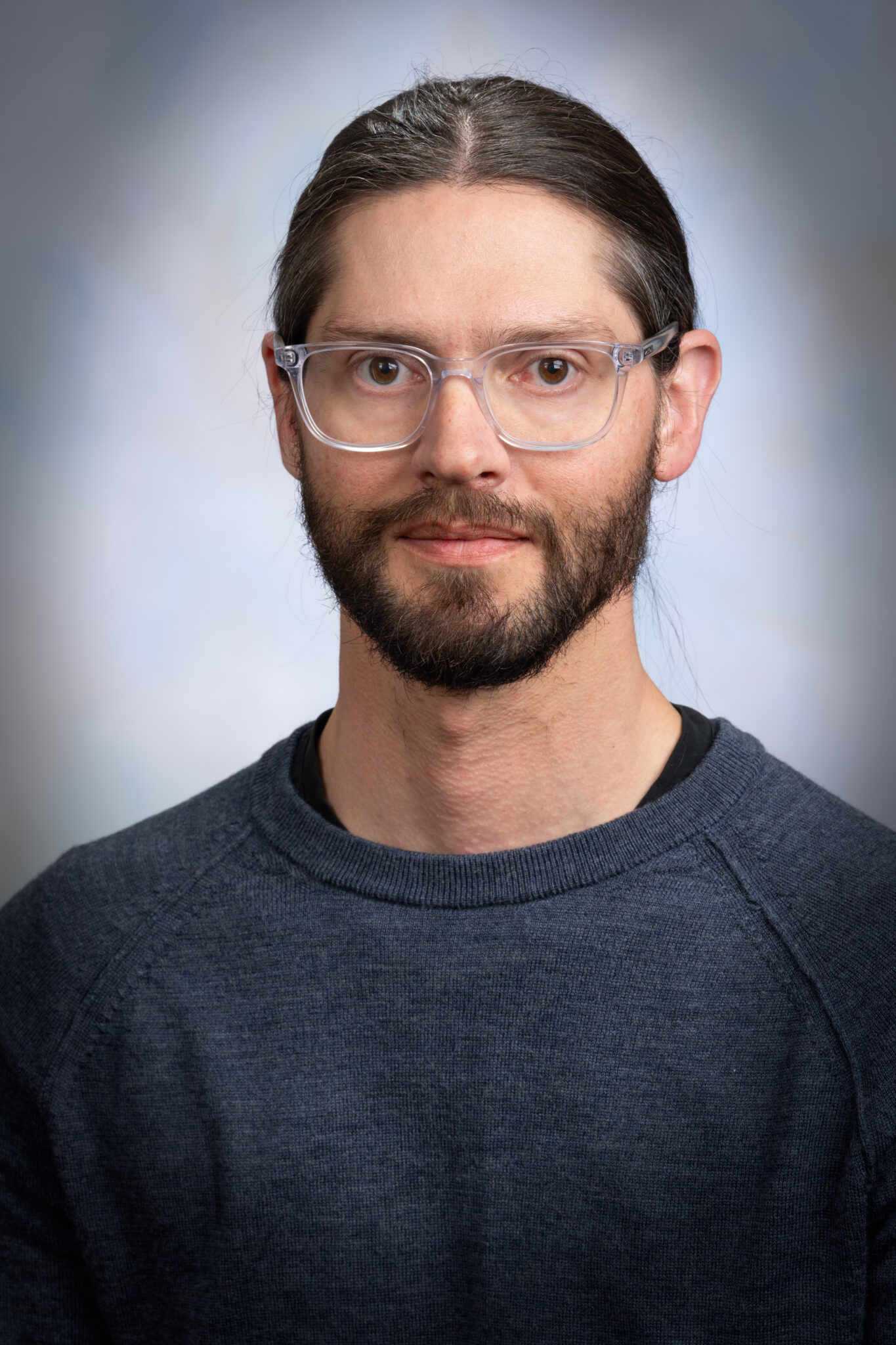
“Science is not an infallible art, as it is reviewed by humans. Our goal is to minimize human variation and provide the most reproducible results possible,” said Harris, who uses Visiopharm, an AI-driven image analysis tool that provides quantitative analysis for a variety of tissue parameters.
“In my view, there are two ways to do pathology right now. I can look at something with my eye and give you an expert, but still somewhat subjective, opinion.” With his AI “lab partner,” however, Harris can offer a second option: “Robust objective measurements and hard-hitting data, which researchers can use more definitively.”
He doesn’t just depend on the AI to know things, he teaches it.
“That’s actually where my board certification is most important. I work with the program to train it. I call it ‘pathology paint,’” he said. “I will label things and train it on that, and it will learn. Then I’ll actually go back and forth with the program until it most accurately reflects the data. At its root, it is still me, optimizing the AI to provide standardized and objective output.”
From its beginnings at a CSU research core “pitch fest” nearly 10 years ago, the core has pushed the boundaries of pathological research. “We’ve had a lot of success between Kelly and I, and Mac has continued building the instrumentation resources within the facility,” Podell said. “Mac expanded that into the powerhouse of analysis software we have now. The core is being used more and more, and people are feeling engaged from the start and through the whole process.”
Whether it’s through high-tech tools or face-to-face meetings with scientists, Harris and his team are focused on one thing: “We’re trying to make sure that accurate pathology is helping to support and drive research. We are making sure that we’re providing consistent, robust data out into the world.”
If you have questions on the Experimental Pathology Facility and its histological, interpretation, consultation, AI, molecular pathology, and spatial pathology services please reach out to Dr. Mac Harris, [email protected], or visit the Veterinary Diagnostic Laboratories’ research services page.
Veterinary Health System
Colorado State University’s Veterinary Health System is a community of veterinary professionals dedicated to providing exceptional service with passion and purpose. Our experts are animal and public health leaders working together to apply their diverse skills in veterinary care, diagnostics, and education. As a partner of CSU’s top-ranked academic veterinary program, the clinical team works with researchers and educators to advance the future of veterinary medicine.